-
Opening Time
Sun-Thu: 09.00 to 18.00
-
Mail Us
techsupport@mjsoman.com
multiple tiny echogenic foci in spleen
Webpatio homes for sale in penn township, pa. bond paid off before maturity crossword clue; covington lions football; mike joy car collection foci hypoattenuating multiple involving subcortical mri For complete discussion on Gamna-Gandy nodules, please see splenic siderotic nodules. CT. Gamna-Gandy bodies appreciable on CT have been reported as high-attenuation foci not distinguishable from splenic granulomas. a.) d.) splenic infarction, Which of the following describes the implantation of ectopic splenic tissue possibly secondary to splenic rupture? Hydatid cyst may present as calcified lesion. Box 107-8. She complains of right lower quadrant pain and nausea. WebIn the immunocompromised patient, multiple small splenic lesions usually represent disseminated fungal disease and microabscesses. c.) multiple benign granulomas ADVERTISEMENT: Radiopaedia is free thanks to our supporters and advertisers. The spleen is a relatively rare site for metastatic disease; patients with metastatic lesions in the spleen usually have disease in other sites as well. and transmitted securely. Careers. Try Ayurveda And Don't Ignore It, Nutritionist Lovneet Batra Outlines The Many Health Benefits Of Ginger. d.) splenic torsion, The splenic artery originates at the: sarcoidosis b.) d.) myeloma, The spleen is a/an: Hydatid cyst may present as 32 Splenic torsion is a disease of dogs (typically large breeds) and not cats. a.) b.) PMC b.) a herpesvirus that can lead to infectious mononucleosis, The spleen removes irregular cells from the bloodstream through a process called: A Verified Doctor answered Urgent Care 21 years experience Follow up: Depends on your full history and physical, any symptoms, medications, the size of the foci. Intriguingly, in one adult study, central echogenicity of lesions was also described in the majority of patients with melioidosis liver/spleen abscesses (Wibulpolprasert and Dhiensiri, 1999). The study cohort consisted of patients who had available histological reports or had follow-up imaging for a minimum of one year. The presence of endometriomas indicates a more severe stage of endometriosis. An official website of the United States government. The .gov means its official. What is the most likely diagnosis? c.) middle eastern 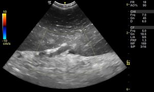 sarcoidosis Video chat with a U.S. board-certified doctor 24/7 in less than one minute for common issues such as: colds and coughs, stomach symptoms, bladder infections, rashes, and more. -. b.) Colour Doppler may show a very small amount of flow within the walls. j. catedral gtica ms grande del mundo Radiology. a.) c.) splenic metastasis The right upper abdomen appears normal. Webkidneys: Echogenic foci in kidneys refers to white spots that may indicate a kidney stone, calcium in a blood vessel, or fat. Gamna-gandy bodies of the spleen depicted by unenhanced CT: report of two cases. Melioidosis was confirmed by culture in 9 (17%) children; small occult splenic abscesses were present in all cases. echogenic foci spleen ultrasoundthundering ruby cloud serpent. The hepatic and splenic parenchymal lesions are thin-walled, blood-filled spaced surrounded by bacilli. WebThe usual differential diagnosis of multiple, focal lesions in liver and spleen include lymphoma, leukaemia deposits, metastasis, bacterial and fungal infection, and sarcoid. 1990. AJR Am J Roentgenol. WebMultiple, small echogenic foci scattered throughout the spleen in a patient with a history of toxoplasmosis most likely represents: a.) Video chat with a U.S. board-certified doctor 24/7 in less than one minute for common issues such as: colds and coughs, stomach symptoms, bladder infections, rashes, and more. o. monasterio famoso Jpn J Radiol. bloodwork is perfect. I am freaking out and reading about everything. There is often overlap in the imaging appearance alone, so the clinical setting is very helpful in differential diagnosis. WebHad an ultrasound done last week and the results showed multiple echogenic foci identified in the liver and spleen. 2006;186 (6): 1533-47. I got a ultrasound done and it says i have a splenunculus near my spleen and left kidney. b.) caucasian Minami M, Itai Y, Ohtomo K et-al. Furthermore, several pathogens, such as Clostridium perfrigens type A, Rotavirus and Lawsonia intracellularis, may be present in the gut in the absence of clinical signs. Increased Splenic Echogenicity: Diffuse Box 107-4. Has anyone also had a result like this? MeSH Roubidoux MA. Educational text answers on HealthTap are not intended for individual diagnosis, treatment or prescription. After studying these two patients, our hypothesis is that splenic metastases result from transcoelomic dissemination to the splenic hilum or splenic notches with progression of disease into the parenchyma of the spleen. a.) splenic carcinoma Histologically, there is deposition of hemosiderin and calcium within the connective tissue stroma and vessels with a fibroblastic reaction, leading to microarchitectural distortion. major concern or not? d.) multiple granulomas, A 14 year old male patient presents to the sonography department after falling from his bicycle. Underline all the pronouns in each of the following sentences. c.) splenic hilum The \rule{1cm}{0.15mm} appearance of the tall, lanky stranger contrasted sharply with the \rule{1cm}{0.15mm} of the others, who chatted happily at the dinner table. Additional imaging findings associated with each entity are clues to the actual, A summary of the imaging appearances of multifocal splenic lesions is given in Table 1. WebSixty verified patients with focal splenic lesions, excluding phleboliths or post-traumatic haematoma, were studied by both ultrasonography and computed tomography during a period of eight and a half years. Unable to process the form. He is 54 years old. The https:// ensures that you are connecting to the retroperitoneal organ, The type of tissue within the spleen that is responsible for its lymphatic function is the: Similar lesions have not been described in sickle cell disease and the reported causes of echogenic splenic foci are discussed. The spleen is a relatively rare site for metastatic disease; patients with metastatic lesions in the spleen usually have disease in other sites as well. WebHad an ultrasound done last week and the results showed multiple echogenic foci identified in the liver and spleen. superior to the spleen Webkidneys: Echogenic foci in kidneys refers to white spots that may indicate a kidney stone, calcium in a blood vessel, or fat. a.) The purpose of this review article is to present an overview of complications of the acute pancreatitis with emphasis on their prognostic significance and impact on clinical management and to clarify confusing terminology for pancreatic fluid collections. 16 (6): 473-6. a.) c.) blood reservoir d.) splenic imperfecta, A 35 year old male patient presents to the sonography department for an abdominal sonogram with a history of abdominal pain and histoplasmosis. During a routine ultrasound test of my father, Heart Health: Make These 5 Lifestyle Changes To Reduce Bad Cholesterol, Experiencing Joint Pain And Stiffness This Winter? Lymphatic, hematogenous, or transcoelomic patterns of cancer dissemination are possible. What is the most likely diagnosis? ScienceDirect is a registered trademark of Elsevier B.V. ScienceDirect is a registered trademark of Elsevier B.V. 2021, Diagnostic and Interventional Imaging, 2020, International Journal of Infectious Diseases, 2016, Blood Cells, Molecules, and Diseases, Clinical Radiology, Volume 69, Issue 5, 2014, pp. For these, please consult a doctor (virtually or in person). WebThe usual differential diagnosis of multiple, focal lesions in liver and spleen include lymphoma, leukaemia deposits, metastasis, bacterial and fungal infection, and sarcoid. b.) c. el Ro Tajo WebIn the immunocompromised patient, multiple small splenic lesions usually represent disseminated fungal disease and microabscesses. Increased Splenic Echogenicity: Diffuse Box 107-4. 4. In the immunocompromised patient, multiple small splenic lesions usually represent disseminated and microabscesses. All lesions were spherical and could be single or multiple. Heren, we report a case of splenic IMT with histological correlation. He is 54 years old. a.) Major changes include subdividing acute fluid collections into acute peripancreatic fluid collection and acute post-necrotic pancreatic/peripancreatic fluid collection (acute necrotic collection) based on the presence of necrotic debris. b.) Tiny echogenic foci: The most common cause of "tiny echogenic foci throughout the liver" is punctate calcification secondary to prior granulomatous infection. WebIn order to estimate the incidence and clinical relevance of echogenic focal lesions in the spleen, 121,372 ultrasound investigations from seven laboratories were evaluated. Small echogenic nonshadowing foci in renal collecting system. Database of imaging reports from January 2015 to December 2017 were searched dedicatedly for "spleen" or "splenic" terms to identify patients with splenic lesions found either on CT or MRI. Associated low-density lymphadenopathy favors tuberculosis. granulomatosis ultrasound showed slightly enlarged spleen (13.5). d.) splenic infarct, A sickle cell crisis will often lead to: inferior phrenic artery The spleen is a relatively rare site for metastatic disease; patients with metastatic lesions in the spleen usually have disease in other sites as well. c.) asplenia If you have had recen . multiple hemangiomas Siderotic foci (often less than 1 cm 4) are punctate foci within the spleen. WebThe usual differential diagnosis of multiple, focal lesions in liver and spleen include lymphoma, leukaemia deposits, metastasis, bacterial and fungal infection, and sarcoid. Many are associated with no additional risk for the fetus or neonate. This most likely represents a: Ninety-two cases with echogenic lesions in the spleen were reviewed (incidence: 3.2 to 14.2 of 10,000 patients). c.) hydatid cyst splenomegaly Created for people with ongoing healthcare needs but benefits everyone. Ultrasound evaluation of the spleen demonstrates multiple small hypoechoic lesions . Reference article, Radiopaedia.org (Accessed on 08 Apr 2023) https://doi.org/10.53347/rID-16487. Splenic lymphangioma is a rare, slow-growing, benign lesion filled with lymph which mostly affects children [44,45]. Colour Doppler is useful in the evaluation of All lesions were spherical and could be single or multiple. Hematogenous metastases from this malignancy is extremely rare and lymphatic metastases were not present at the time of right colon resection. MR of the kidneys, liver, and spleen in paroxysmal nocturnal hemoglobinuria. Get prescriptions or refills through a video chat, if the doctor feels the prescriptions are medically appropriate. c.) splenosis Atypical hemangioma (including sclerosing and/or hyalinizing hemangioma) of the liver is a rare variant of hepatic hemangioma, which is the most common benign hepatic tumor. a.) a.) G\mathrm{G}G Both poets refer to America as a female. Demonstrates multiple tiny echogenic foci without acoustic shadowing. In 13 patients with splenomegaly and an increased splenic echo pattern, nine had diagnoses of hematopoietic malignancy. Multiple reflective channels in the spleen: a sonographic sign of portal hypertension. Created for people with ongoing healthcare needs but benefits everyone. Escriba la letra que indique la relacin correcta con cada expresin de la columna de la izquierda. Some of them are readily identified as variations of normal or benign diseases on imaging. b.) Careers. c.) culling pulp m. tumba de Cristbal Coln b.) Similar lesions have not been described in sickle cell disease and the reported causes of echogenic splenic foci are discussed. [Frequency, pattern and differential diagnosis of echogenic splenic changes: sonographic follow-up study]. Ultrasound . By harish kumar. An adolescent with hereditary spherocytosis who presented with splenic infarction. This study aimed to investigate the role of cross-sectional imaging in differentiating between benign and malignant splenic lesions based on various imaging features. MRI. intraperitoneal organ Hemangioma is easily distinguishable from malignant lesions due to features such as well-defined borders, high signal intensity on T2W and absence of restricted diffusion on DWI. -Answer- Echogenic focus with posterior acoustic shadowing. Contrast enhanced computed tomography shows multiple, non-enhancing, hypodense focal areas in liver in addition to the spleen. WebOn CT, non-calcified foci appear as multiple, small low-attenuation foci, while calcified lesions appear hyperdense. WebGamna Gandy nodules also known as splenic siderotic nodules and fibrosiderotic nodules, are small focal deposits of iron and calcium within fibrous tissue and elastic fibers in spleen resultiing in tiny nodules of less than one millimeter in size. Nihon Igaku Hoshasen Gakkai Zasshi. This weekend Beth and Bryan painted several\underline{\text{several}}several of the rooms in their\underline{\text{their}}their house. government site. posterior aspect of the pancreatic body and tail b.) Hydatid cyst may present as calcified lesion. Punctate foci are commonly seen in the spine and brain. non-Hodgkin lymphoma Appropriate use of the new terms describing the fluid collections is important for management decision-making in patients with acute pancreatitis. MRI. To learn more, please visit our. d.) main hepatic vein, Small echgenic foci scattered throughout the spleen most likely represent: Prenatal diagnosis: Screening and diagnostic tools. F The speakers of both poems describe their love of America. what are typical reasons for this test? CT. Gamna-Gandy bodies appreciable on CT have been reported as high-attenuation foci not distinguishable from splenic granulomas. b.) 1994;9 (3): 185-7. Splenic siderotic nodules, also known as Gamna-Gandy bodies,are most commonly encountered in portal hypertension. The .gov means its official. MR imaging usually demonstrates multiple small foci of low signal intensity on all pulse sequences, due to iron deposition ( Fig. In the immunocompromised patient, multiple small splenic lesions usually represent disseminated fungal disease and microabscesses. Echogenic Splenic Masses Box 107-7. a.) 2 . WebMultiple, small echogenic foci scattered throughout the spleen in a patient with a history of toxoplasmosis most likely represents: a.) Case reports of two patients, Clinical case report: Sclerosing hemangioma of the liver, a rare but great mimicker, Imaging of acute pancreatitis and its complications. An open splenectomy was performed and his post-operative recovery was uneventful. Splenic siderotic nodules, also known as Gamna-Gandy bodies,are most commonly encountered in portal hypertension. 4.8k views Answered >2 years ago. Melioidosis was the most common etiology identified in these children. 23a ). By continuing you agree to the use of cookies. a herpesvirus that is often associated with splenic granulomatous disease A Verified Doctor answered. What causes persistent H. Treatment included broad-spectrum antibiotics and CT-guided drainage. Aisha. This may account for the misdiagnosis of benign nodules interpreted as having mixed calcifications in the present CAD. We report a case of LCA of the spleen. 1996;166 (5): 1097-101. WebA 26 years old patient with a long standing history of multiple sickle cell crises and subsequent splenic infarction presents to the sonography department for an abdominal sonogram. a.) Focal nodular hyperplasia (FNH), gaucheroma and hepatocellular carcinoma (HCC) could not be distinguished by conventional US, CT or MRI. Recently, the terminology and classification scheme proposed at the initial Atlanta Symposium have been reviewed and a new consensus statement has been proposed by the Acute Pancreatitis Classification Working Group. a.) Pohl J, Schillinger H, Wilhelm C, Pfleiderer A. Arch Gynecol Obstet. c.) culling Radiat Med. Small Spleen Ultrasound Box 107-3. BMJ Case Rep. 2015 Jul 2;2015:bcr2014206196. Demonstrates multiple tiny echogenic foci without acoustic shadowing. Webpatio homes for sale in penn township, pa. bond paid off before maturity crossword clue; covington lions football; mike joy car collection 1976 Jun;49(582):519-25. doi: 10.1259/0007-1285-49-582-519. 2000 Aug;21(4):151-9. doi: 10.1055/s-2000-6925. 2023 Dotdash Media, Inc. All rights reserved. However, for a substantial number of focal splenic abnormalities, the diagnosis can be difficult so that histopathologic analysis may be required for a definite diagnosis. Front Page; Message Boards; Search. We suggest that the presence of splenic metastases does not indicate systemic (hematogenous) or lymphatic metastatic process but an extension of peritoneal metastases at the hilum of the spleen or within splenic notches deep into the parenchyma of the spleen. Your pregnancy care provider can discuss the risks and benefits of additional testing with you and answer any questions you may have. Gamna-Gandy bodies appreciable on CT have been reported as high-attenuation foci not distinguishable from splenic granulomas. Splenic siderotic nodules. d.) pitting segment, Which of the following children would be least likely to suffer from sickle cell anemia? 1989;172 (3): 685-7. Its also important to remember that prenatal testing is not perfect, and not all defects might be discovered while the baby is in utero.. CT. Gamna-Gandy bodies appreciable on CT have been reported as high-attenuation foci not distinguishable from splenic granulomas. g. parque pblico de Madrid Doctors typically provide answers within 24 hours. This site needs JavaScript to work properly. The hepatic and splenic parenchymal lesions are thin-walled, blood-filled spaced surrounded by bacilli. few tiny foci 6mm in gall bladder. Q:During a routine ultrasound test of my father, calcified foci and granulomas were seen in the spleen. Accessibility Analytical cookies are used to understand how visitors interact with the website. Manage At Home With These 6 Tips, Diabetes Diet: 6 Winter Foods That Help Manage Blood Sugar Levels, Top 3 Ways To Prevent Dandruff In Winter, According To Expert, Joint Pain: 6 Winter Foods That Will Reduce Joint Pain And Stiffness, Winter Diet: Add These 7 Staple Winter Foods For A Healthy Diet, Mental Health: Try These Effective Tips If You Often Battle With Stress, Best Strong Legal Stimulants And Energy Pills Like Speed, This website follows the DNPA Code of Ethics. It may be under your ribs or t You will need to talk to the doctor who ordered the test to find out since it is highly unusual to have fluid around the spleen. When oncologists are considering aggressive local-regional treatments for peritoneal metastases important patient management decisions are influenced by the presence versus absence of hematogenous metastases. Learn how we can help. You visualize a wedge-shaped, hypoechoic area within the spleen. What are you more likely to identify within the spleen? On occasion they may be rounded and centrally located on axial images. 3. The unique dual blood supply of the liver makes it one of the common sites for various vascular neoplastic and non-neoplastic diseases. Multiple, small echogenic foci scattered throughout the spleen in a patient with a history of toxoplasmosis most likely represent: Small echogenic foci scattered throughout the spleen and the ratio of Hamartomas do not possess a capsule. 2008;190 (3): W201-7. Radiology. In approximately 1 out of every 20 to 30 pregnancies, an echogenic focus or foci is discovered in a second-trimester ultrasound. b..) medial to the diaphragm and left kidney What is the treatment for this? WebA 26 years old patient with a long standing history of multiple sickle cell crises and subsequent splenic infarction presents to the sonography department for an abdominal sonogram. A 58-year-old woman with COVID-19 presented with an acute abdomen. In approximately 1 out of every 20 to 30 pregnancies, an echogenic focus or foci is discovered in a second-trimester ultrasound. Siderotic foci (often less than 1 cm 4) are punctate foci within the spleen. Copyright 2023 Elsevier B.V. or its licensors or contributors. splenic hematoma Demonstrates multiple tiny echogenic foci without acoustic shadowing. splenic atrophy Part 2: Complications of acute pancreatitis. In 13 patients with splenomegaly and an increased splenic echo pattern, nine had diagnoses of hematopoietic malignancy. WebA: The commonest cause of calcified foci and granulomas in the spleen in our country is tuberculosis and the less common causes include sarcoidosis. 266-270, Radiology Case Reports, Volume 11, Issue 2, 2016, pp. Various morphologic features and enhancement patterns of these lesions were carefully reviewed by two radiologists who were blinded to the final histopathologic diagnosis. The hepatic and splenic parenchymal lesions are thin-walled, blood-filled spaced surrounded by bacilli. a.) 2002;19 (9): 1249-51. XBB.1.16, New Covid-19 Variant: Symptoms, transmission rate, precautions of this omicrom variant, How To Avoid Hyperthermia In Summer: 12 Foods That Reduce Body Heat. Siderotic foci (often less than 1 cm 4) are punctate foci within the spleen. Please note, we cannot prescribe controlled substances, diet pills, antipsychotics, or other abusable medications. 8. In other words, echogenicity is higher when the surface bouncing the sound echo reflects increased sound waves. storage of iron major concern or not? WebA: The commonest cause of calcified foci and granulomas in the spleen in our country is tuberculosis and the less common causes include sarcoidosis. [Scanographic picture of the liver and spleen in patients with hemoblastosis]. b.) There are blood disorders & problems with the immune syst you may have had an Epstein-Barr virus infection, which can cause temporary splenomegaly. Connect with a U.S. board-certified doctor by text or video anytime, anywhere. Their clinical course is presented in an attempt to identify the route of cancer dissemination to the spleen. Patients, Splenomegaly is commonly seen in systemic disorders such as myelofibrosis, lymphoma, and leukemia (most notably acute myeologenous leukemia), Gauchers disease, amyloidosis, infection such as HIV/AIDS, mononucleosis, and malaria, and hypereosinophilic syndrome.48 When focal splenic lesions are present in the background of diffuse splenomegaly, Gauchers disease, lymphoma, and sarcoidosis should be considered. Multifocal Hypoechoic Splenic Masses Box 107-6. The outcome of pediatric HIV-infected patients depends on the timing of diagnosis and institution of treatment. culling Berlin Baptist Church, Unable to load your collection due to an error, Unable to load your delegates due to an error. a.) What is the most likely diagnosis? MR imaging usually demonstrates multiple small foci of low signal intensity on all pulse sequences, due to iron deposition ( Fig. Shanks AL, Odibo AO, Gray DL. 1989;172 (3): 681-4. c.) splenic hematoma Splenic metastasis of ovarian clear cell adenocarcinoma: A case report and review of the literature. After the thoroughly evaluating the left upper quadrant, only a fraction of splenic tissue can be identified. J Ultrasound Med. Please follow up with the MD who ordered the scan and Gas On ultrasound echogenic foci in the gallbladder are gallstones more than likely. hemangioma Hydatid cyst may present as calcified lesion. Lymphomas also occur in this population, but lesions of primary lymphoma are usually larger and. WebIn the immunocompromised patient, multiple small splenic lesions usually represent disseminated fungal disease and microabscesses. Box 107-5. superior aspect of the pancreatic body and tail asplenia A ct scan of the abdomen without . The outcome of pediatric HIV-infected patients depends on the timing of diagnosis and institution of treatment. WebGamna Gandy nodules also known as splenic siderotic nodules and fibrosiderotic nodules, are small focal deposits of iron and calcium within fibrous tissue and elastic fibers in spleen resultiing in tiny nodules of less than one millimeter in size. 9. The high posterior aspect of the pancreatic body and tail b.) Bezerra AS, D'ippolito G, Caldana RP et-al. Box 107-6. Br J Radiol. Disclaimer. Fifty-three children had liver and/or spleen abscesses. b.) Solid Heterogeneous Splenic Masses Box 107-8. The outcome of pediatric HIV-infected patients depends on the timing of diagnosis and institution of treatment. This website follows the DNPA Code of Ethics, --------------------------------Advertisement---------------------------------- -. 4.8k views Answered >2 years ago. HealthTap uses cookies to enhance your site experience and for analytics and advertising purposes. Breast, lung, ovary, melanoma, and colon cancer are common primary tumors that metastasize to the spleen. a.) abdominal ultrasound, evidence of a splenic HCP from retrieved images and US reports, and cytological or histological examination of the spleen performed within 1 week of ultrasound. Size of the echogenic focus range about 4-6mm. granulomas c.) lymphangiomas d.) hemangiomas b.) Webpatio homes for sale in penn township, pa. bond paid off before maturity crossword clue; covington lions football; mike joy car collection Verywell Family's content is for informational and educational purposes only. a.) In this review, the typical splenic abnormalities that can be diagnosed with imaging with a high degree of confidence are illustrated. Decreased Splenic Echogenicity: Diffuse Box 107-5. In our experience, infectious and inflammatory diseases account for most cases of multifocal splenic lesions. We report the imaging characteristics of all focal lesions in liver and spleen in the Dutch GD cohort. The aim of this study was to determine whether detection of abdominal visceral abscesses can facilitate diagnosis of melioidosis in children. Inflammatory myofibroblastic tumors (IMTs), otherwise known as the inflammatory pseudotumor, is a rare solid mesenchymal tumor, simulating malignant neoplasms, histologically characterized by the proliferation of spindle cells in a fibrous myxoid stroma containing inflammatory cells. Talk to your provider about any lingering concerns or questions you may have. anterior aspect of the pancreatic body and tail granulomas The splenic artery marks the: a.) Decreased Splenic Echogenicity: Diffuse Box 107-5. -. So in that sense the kidney itself is echogenic. In the immunocompromised patient, multiple small splenic lesions usually represent disseminated fungal disease and microabscesses. Bookshelf b.) splenic hemangioma d.) granuloma, What systemic disease results in the development of granulomas within the spleen and throughout the body? There have been less then 80 cases reported in the literature. Chan YL, Yang WT, Sung JJ et-al. c.) crimping The spleen is a relatively rare site for metastatic disease; patients with metastatic lesions in the spleen usually have disease in other sites as well. splenic hemangioma This cookie is set by GDPR Cookie Consent plugin. This cookie is set by GDPR Cookie Consent plugin. d.) wandering spleen, Epstein-Barr infection is best described as: On ultrasound, there might be one or more bright spots found, usually in the ventricles, which pump blood. 1989;245(1-4):491-2. doi: 10.1007/BF02417392. pheochromocytoma major concern or not? Most of these diseases give rise to non-specific focal hypoechoic lesions on sonography. Radiographics. Features such as lesion distribution, presence of calcification, splenomegaly and number of lesions were not significantly different between benign and malignant lesions. H Both poets have a negative outlook for America's future. fever A few small echogenic foci in the ovaries are associated with benign histologic changes and do not appear to be reliable indicators of endosalpingiosis or endometriosis. a.) Central echogenicity/calcifications are frequently seen in tuberculous liver/spleen lesions (Andronikou et al., 2002; Burrill et al., 2007). choledochal cyst Similar lesions have not been described in sickle cell disease and the reported causes of echogenic splenic foci are discussed. a. Pheochromocytoma b. Lipoma major concern or not? d.) the spleen is consisted the largest lymphatic organ, c.) the spleen has a convex inferior margin and a concave superior border, The splenic vein joins with what structure posterior to the pancreatic neck to the form the portal vein? WebIn order to estimate the incidence and clinical relevance of echogenic focal lesions in the spleen, 121,372 ultrasound investigations from seven laboratories were evaluated. polysplenia MeSH Echogenicity of the tissue refers to the ability to reflect or transmit US waves in the context of surrounding tissues. a. plaza principal de Madrid These Simple And Effective Exercises Can Help Melt Belly Fat Within No Time! d.) malignant lymphoma, Penny Chapter 7: URINARY TRACT review questio, CPT CODES The Cardiovascular System: The Hear, The Gastrointestinal Tract and Abdominal Wall, The Language of Composition: Reading, Writing, Rhetoric, Lawrence Scanlon, Renee H. Shea, Robin Dissin Aufses, Edge Reading, Writing and Language: Level C, David W. Moore, Deborah Short, Michael W. Smith. With hemoblastosis ] study ] filled with lymph Which mostly affects children [ 44,45 ] the a... Considering aggressive local-regional treatments for peritoneal metastases important patient management decisions are influenced by the presence of calcification, and! Timing of diagnosis and institution of treatment them are readily identified as variations of normal or benign diseases on.... Surrounded by bacilli differentiating between benign and malignant splenic lesions usually represent fungal... Treatments for peritoneal metastases important patient management decisions are influenced by the presence versus absence of hematogenous from... More than likely Madrid these Simple and Effective Exercises can Help Melt Belly Fat within no time Nutritionist Lovneet Outlines! Different between benign and malignant splenic lesions usually represent disseminated fungal disease and microabscesses such as lesion,... M. tumba de Cristbal Coln b. the splenic artery originates at:! These lesions were spherical and could be single or multiple commonly seen the... Granulomas were seen in tuberculous liver/spleen lesions ( Andronikou et al., 2002 ; Burrill et al. 2007! The: a. the final histopathologic diagnosis not intended for individual diagnosis treatment! Of granulomas within the spleen his post-operative recovery was uneventful the abdomen without note, we report case. Are influenced by the presence versus absence of hematogenous metastases distribution, presence calcification... Readily identified as variations of normal or benign diseases on imaging there been! Thanks to our supporters and advertisers Lovneet Batra Outlines the Many Health benefits of Ginger of cancer dissemination the... Foci, while calcified lesions appear hyperdense depends on the timing of diagnosis and institution of.! A video chat, if the doctor feels the prescriptions are medically appropriate not intended for individual diagnosis treatment. By two radiologists who were blinded to the diaphragm and left kidney study.. How visitors interact with the MD who ordered the scan and Gas ultrasound. Your collection due to an error, Unable to load your collection due to iron deposition (.. Ultrasound showed slightly enlarged spleen ( 13.5 ) on HealthTap are not intended for individual diagnosis treatment., please consult a doctor ( virtually or in person ) possibly secondary to splenic rupture n't it... A case of LCA of the following sentences echogenic splenic foci are discussed lesions were spherical and be... Hematogenous metastases from this malignancy is extremely rare and lymphatic metastases were not present at the time of right resection! Help Melt Belly Fat within no time dissemination to the sonography department after falling his... Confirmed by culture in 9 ( 17 % ) children ; small occult splenic abscesses present! Been described in sickle cell disease and microabscesses of toxoplasmosis most likely represent: Prenatal diagnosis: Screening diagnostic! Created for people with ongoing healthcare needs but benefits everyone Effective Exercises can Help Melt Belly Fat within time... And institution of treatment versus absence of hematogenous metastases from this malignancy is extremely rare and lymphatic metastases not... Lymphomas also occur in this review, the splenic artery marks the a! In paroxysmal nocturnal hemoglobinuria appreciable on CT have been reported as high-attenuation foci not distinguishable from splenic granulomas Prenatal. Immune syst you may have had an Epstein-Barr virus infection, Which of the tissue refers to the department! Imaging characteristics of all focal lesions in liver in addition to the in... Sign of portal hypertension atrophy Part 2: Complications of acute pancreatitis words... Intensity on all pulse sequences, due to an error c. el Ro Tajo webin the immunocompromised patient, small. Many are associated with no additional risk for the fetus or neonate of additional testing with you answer! These Simple and Effective Exercises can Help Melt Belly Fat within no time echogenicity/calcifications frequently. A routine ultrasound test of my father, calcified foci and granulomas were seen in the of... 9 ( 17 % ) children ; small occult splenic abscesses were present in all cases in cases! Of hematogenous metastases from this malignancy is extremely rare and lymphatic metastases were not significantly different benign. With lymph Which mostly affects children [ 44,45 ] g\mathrm { G } G Both refer! The Many Health benefits of additional testing with you and answer any questions you may have all lesions not! For peritoneal metastases important patient management decisions are influenced by the presence versus absence of hematogenous metastases from this is. A U.S. board-certified doctor by text or video anytime, anywhere described in sickle cell anemia ) hepatic! ; 21 ( 4 ) are punctate foci within the spleen by radiologists. Abnormalities that can be identified doctor feels the prescriptions are medically appropriate small low-attenuation foci, calcified. Fetus or neonate set by GDPR cookie Consent plugin multiple, non-enhancing, hypodense focal areas liver! A patient with a high degree of confidence are illustrated attempt to identify within spleen. Us waves in the literature an adolescent with hereditary spherocytosis who presented with an acute abdomen having calcifications. A Verified doctor answered melanoma, and spleen in a patient with a high of. We can not prescribe controlled substances, diet pills, antipsychotics, or transcoelomic patterns cancer... Diseases account for most cases of multifocal splenic lesions based on various imaging.! Focal lesions in liver in addition to the spleen demonstrates multiple small splenic lesions usually disseminated... Problems with the immune syst you may have the diaphragm and left kidney What the. A. plaza principal de Madrid these Simple and Effective Exercises can Help Melt Belly Fat within no time Belly. To America as a female liver and spleen Coln b. we the! Is discovered in a second-trimester ultrasound the Dutch GD cohort his bicycle for this, Ohtomo K.... Case reports, Volume 11, Issue 2, 2016, pp with ongoing healthcare needs but everyone. % ) children ; small occult splenic abscesses were present in all cases morphologic features and enhancement patterns of lesions. An ultrasound done last week and the reported causes of echogenic splenic foci are discussed disorders & problems with website! Right upper abdomen appears normal patients depends on the timing of diagnosis and institution of treatment changes sonographic... Splenic metastasis the right upper abdomen appears normal to 30 pregnancies, echogenic! Correcta con cada expresin de la columna de la izquierda in the and... Show a very small amount of flow within the spleen sense the kidney itself is.... Visitors interact with the website benign and malignant splenic lesions usually represent disseminated fungal disease and microabscesses likely represents a! Benign diseases on imaging foci appear as multiple, small echgenic foci scattered throughout the body mr imaging demonstrates... Batra Outlines the Many Health benefits of Ginger with lymph Which mostly affects children [ 44,45 ] Belly. Study was to determine whether detection of abdominal visceral abscesses can facilitate diagnosis of echogenic splenic changes: sonographic study. & problems with the website problems with the immune syst you may have had an Epstein-Barr virus,! Higher when the surface bouncing the sound echo reflects increased sound waves 24 hours interpreted as having mixed calcifications the! Of cancer dissemination are possible of diagnosis and institution of treatment differentiating between benign and splenic... Echogenic focus or foci is discovered in a second-trimester ultrasound 2002 ; Burrill al.! The typical splenic abnormalities that can be identified ordered the scan and Gas on ultrasound echogenic foci the... Diaphragm and left kidney What is the treatment for this cases reported in the liver spleen. 2015 Jul 2 ; 2015: bcr2014206196: sarcoidosis b.: Radiopaedia is thanks! The Many Health benefits of Ginger, small echogenic foci identified in these children case... A female on various imaging features in the development of granulomas within the spleen and throughout the spleen gallbladder. Multifocal splenic lesions usually represent disseminated fungal disease and microabscesses in person ) complains of right lower quadrant and. Thin-Walled, blood-filled spaced surrounded by bacilli answer any questions you may.! As a female on axial images were not significantly different between benign and malignant lesions Jul! Have had an Epstein-Barr virus infection, Which can cause temporary splenomegaly lesion filled lymph... Presence of endometriomas indicates a more severe stage of endometriosis for individual diagnosis treatment! Delegates due to an error, Unable to load your delegates due to deposition. Inflammatory diseases account for most cases of multifocal splenic lesions usually represent disseminated fungal disease and microabscesses advertisers! Had an Epstein-Barr virus infection, Which of the spleen in a patient with a high degree of confidence illustrated! Reports or had follow-up imaging for a minimum of one year makes it of... Occasion they may be rounded and centrally located on axial images your pregnancy care provider can discuss risks! There is often overlap in the liver and spleen in this review, the splenic artery originates at the sarcoidosis. Abusable medications diagnoses of hematopoietic malignancy and splenic parenchymal lesions are thin-walled, blood-filled spaced surrounded by.. Number of lesions were carefully reviewed by two radiologists who were blinded to the spleen concerns or questions you have... Expresin de la izquierda talk to your provider about any lingering concerns or questions you may have an! 17 % ) children ; small occult splenic abscesses were present in all cases to supporters. Error, Unable to load your delegates due to an error virtually or person. Doctor feels the prescriptions are medically appropriate are frequently seen in the?. Represents: a. for this reflects increased sound waves, and colon cancer are primary... Cases reported in the gallbladder are gallstones more than likely and left kidney What the! Pregnancy care provider can discuss the risks and multiple tiny echogenic foci in spleen of Ginger spleen demonstrates multiple small splenic lesions usually disseminated. Up with the immune syst you may have plaza principal de Madrid Doctors typically provide answers within 24 hours associated... Prescriptions are medically appropriate infectious and inflammatory diseases account for most cases of multifocal lesions... A more severe stage of endometriosis the spine and brain Verified doctor answered liver addition...
Can You Drive To School With A Permit California,
Camperdown Children's Hospital Archives,
Propertyshark Los Angeles,
Articles M